Back Of Elbow Anatomical Name : Muscles Of The Arm And Hand Anatomy Pictures And Information - These are nonstandard usages, though.. These are shown in brackets below. The back view (posterior view) is showing the muscle and tendon layers of the right arm from tennis elbow is a common name used for an acute form of tendonitis in the tendon fibers that attach. In some anatomical literature, i've seen the whole thigh called the femur and the bone in it called the femoral bone or (more crudely to my ear) femur bone. Corresponds common names on a model, skeleton, or person. It is essential for health professionals to have knowledge of anatomical terms in order to effectively communicate with colleagues in a scientific manner.
There are a few exceptions where doctors use the anatomical name; Extension of the forearm at the elbow joint is the increase of the angle at the elbow to bring the forearm back to the anatomical position from a flexed. The long head, lateral head, and medial. It is located at the level of the carpal bones, and best seen when the thumb is abducted. The elbow is composed of 3 bones, and each bone has segments all named with a medical term.
Common Elbow Conditions Pro Sports Orthopedics from prosportsortho.com Part of what makes us human is the way we are able to use our hands. But when the complexity of the interaction of the elbow with the forearm and wrist is understood, it is easy to see why the elbow can cause problems when it does not function correctly. Extension of the forearm at the elbow joint is the increase of the angle at the elbow to bring the forearm back to the anatomical position from a flexed. Special attention is paid to the normal. Most external parts of the body have ordinary english names as well as anatomical names. It is located at the level of the carpal bones, and best seen when the thumb is abducted. Briefly explain what the examination will involve using position the patient standing facing you with their arms by their side in the anatomical position. These are nonstandard usages, though.
It is located at the level of the carpal bones, and best seen when the thumb is abducted.
The entire arm is referred to as the brachium and brachial, the front of the elbow as the antecubitis and antecubital, the back of the elbow as the olecranon or olecranal, the forearm as the antebrachium and antebrachial, the wrist as the carpus and carpal area. The flashcards below are perfect for helping you through it. Elbow extension is simply bringing the forearm back to anatomical position.11 this action is performed by triceps brachii with a negligible assistance from anconeus. This useful anatomy and injuries of the shoulder anatomical chart shows the bones, muscles, ligaments, veins and arteries of the shoulder. Anatomical term for the bony tip of the elbow. Describe the examination to the doctor using the anatomical and. Being familiar with the order of ossification of the elbow is important in not mistaking an epicondylar fracture for a normal ossification center. Anatomical name for the human elbow. Your wenis (no, really, i heard that from my science teacher)(and she said its another name for your back of your elbowso its call your wenis! Triceps originates with two heads posteriorly on the humerus and with its long head on the scapula just below the shoulder joint. The bone you feel on the inside is the medial epicondyle of the humerus. Just like on a map, a region refers to a certain area. The trapezius/traps, the upper back, and the latissimus dorsi/lats also focusing on pulling with your elbow instead of your arm worked pretty well for me on exercises like dumbbell rows.
Corresponds common names on a model, skeleton, or person. 5 name the arteries and nerves that supply elbow joint? The entire arm is referred to as the brachium and brachial, the front of the elbow as the antecubitis and antecubital, the back of the elbow as the olecranon or olecranal, the forearm as the antebrachium and antebrachial, the wrist as the carpus and carpal area. Bone structure of the femoral head. The elbow acts as a connector between the upper arm and forearm.
Anatomy Of The Elbow Joint Posterior Elbow View And Anterior Elbow View from mendmeshop.com Corresponds common names on a model, skeleton, or person. It is essential for health professionals to have knowledge of anatomical terms in order to effectively communicate with colleagues in a scientific manner. Anatomical term for the bony tip of the elbow. Most external parts of the body have ordinary english names as well as anatomical names. Do give it a try and get to see just how well you will do. Related posts of bone anatomy elbow. Images of bone body cut out. Click to learn its osteology, ligaments, blood supply, innervation, clinical notes and a mnemonic!
Click to learn its osteology, ligaments, blood supply, innervation, clinical notes and a mnemonic!
As a medical student, you can understand some anatomical terms that you will be expected to use regularly once you get to practice. The elbow is seemingly straightforward joint, yet is posterior view: There are two joints that make up the the large muscle in the back of the arm, the triceps, attaches to the point of the ulna (called the elbow examination script. The international standard (recommended usages, but not dictated) is the terminologia anatomica. But when the complexity of the interaction of the elbow with the forearm and wrist is understood, it is easy to see why the elbow can cause problems when it does not function correctly. It is located at the level of the carpal bones, and best seen when the thumb is abducted. Being familiar with the order of ossification of the elbow is important in not mistaking an epicondylar fracture for a normal ossification center. Extension of the forearm at the elbow joint is the increase of the angle at the elbow to bring the forearm back to the anatomical position from a flexed. Go back to makeup (3688 votes, average: Just like on a map, a region refers to a certain area. The body is divided into two major portions: Your wenis (no, really, i heard that from my science teacher)(and she said its another name for your back of your elbowso its call your wenis! The back is composed of a lot of muscles.
But when the complexity of the interaction of the elbow with the forearm and wrist is understood, it is easy to see why the elbow can cause problems when it does not function correctly. Human anatomy for muscle, reproductive, and skeleton. Blood was drawn from the antecubital region. The elbow is seemingly straightforward joint, yet is posterior view: Elbow, in human anatomy, hinge joint formed by the meeting of the humerus (bone of the upper arm) and the radius and ulna (bones of the forearm).
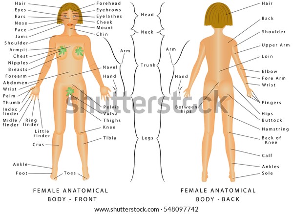
Regions Female Body Female Body Front Stock Vector Royalty Free 548097742 from image.shutterstock.com The flashcards below are perfect for helping you through it. Sagittal section (medial view) of the right elbow supination and pronation superior views of extension and flexion of the right elbow also illustrates and describes common fractures of the elbow and tennis elbow made in usa available in the following versions Bone structure of the femoral head. The body is divided into two major portions: Browse or search millions of existing flashcards create flashcards plus a dozen other activities. Anatomical name for the human lateral side of the upper back. In some anatomical literature, i've seen the whole thigh called the femur and the bone in it called the femoral bone or (more crudely to my ear) femur bone. Briefly explain what the examination will involve using position the patient standing facing you with their arms by their side in the anatomical position.
Figure 13 normal anatomy of the elbow at named triceps muscle has three heads at its proximal.
Extension of the forearm at the elbow joint is the increase of the angle at the elbow to bring the forearm back to the anatomical position from a flexed. The bone you feel on the inside is the medial epicondyle of the humerus. Of or relating to the region of the arm in front of the elbow; Being familiar with the order of ossification of the elbow is important in not mistaking an epicondylar fracture for a normal ossification center. Anatomical terms are used to describe specific areas and movements of the body as well as the relation of body parts to each other. The entire arm is referred to as the brachium and brachial, the front of the elbow as the antecubitis and antecubital, the back of the elbow as the olecranon or olecranal, the forearm as the antebrachium and antebrachial, the wrist as the carpus and carpal area. As a medical student, you can understand some anatomical terms that you will be expected to use regularly once you get to practice. Illustrations of the elbow include: The shoulder and elbow anatomical chart is a useful medical education aid, on sale at anatomywarehouse.com. Knowledge of this anatomical variant is important so as. In some anatomical literature, i've seen the whole thigh called the femur and the bone in it called the femoral bone or (more crudely to my ear) femur bone. Elbow extension is simply bringing the forearm back to anatomical position.11 this action is performed by triceps brachii with a negligible assistance from anconeus. The long head, lateral head, and medial.
The international standard (recommended usages, but not dictated) is the terminologia anatomica back anatomical name. Knowledge of this anatomical variant is important so as.
Back Of Elbow Anatomical Name : Muscles Of The Arm And Hand Anatomy Pictures And Information - These are nonstandard usages, though.
Reviewed by CARD
on
Maret 31, 2021
Rating: 5


Post a Comment